sale.psgmedical@gmail.com
0765.28.48.68


The VISICOIL™ MR magnetic resonance imaging marker system represents a significant advancement in soft tissue tumor localization, providing crucial support to medical teams in planning and implementing precise radiotherapy. Thanks to its advanced multimodal structure, this new generation of markers ensures high-contrast visibility on both MRI and CT scans, optimizing the effectiveness of integrated MR/CT imaging and image-guided radiotherapy (IGRT).
Made entirely from pure platinum, VISICOIL™ MR possesses excellent biocompatibility and delivers sharp, clear images with minimal noise across MR, CT, and ultrasound imaging modalities.
Product Specifications
➔ [BẤM VÀO ĐÂY ĐỂ TẢI VISICOIL FIDUCIAL MARKER CATALOG (PDF)
Discover VISICOIL MR imaging markers for combined MRI & CT imaging
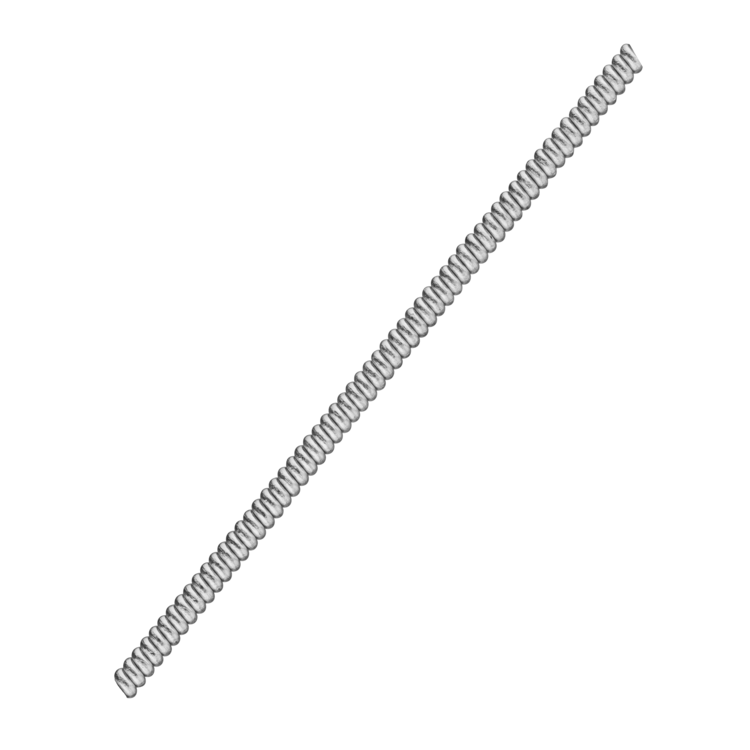
Visicoil MR is made from 99.95% pure platinum, providing clear visibility in both CT and MRI images.
.png)
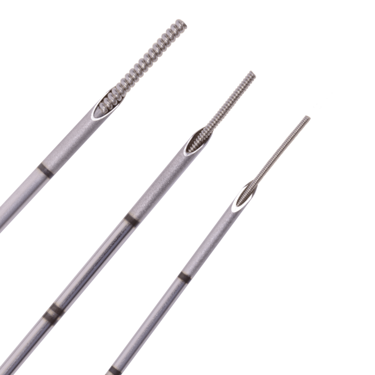
VISICOIL MR measures 0.75 mm x 0.5 cm (implanted for use with the CyberKnife system).
CT scan on GE Discovery CT with a slice thickness of 1.25 mm/slice, a total of 512 slices, no tilt, no overlap, and no gaps between slices.
MRI scan on GE MR750W 3.0T with a slice thickness of 1.3 mm/slice, approximately 180 slices, no overlap, and no gaps between slices. The 3.0 Tesla magnetic field scanner performs pulse sequences (imaging techniques) including:
VISICOIL & VISICOIL MR help reduce image artifacts on CT scans.
Many hospitals still encounter significant image artifacts on CT scans caused by localization markers. This can obscure the boundaries of tumors or target tissues, affecting assessment and treatment planning.
The VISICOIL/VISICOIL MR product line is available in various sizes, allowing physicians to choose the marker best suited to each indication and intervention technique.
VISICOIL and VISICOIL MR are compatible with the following imaging and radiotherapy modalities:
.png)
See It.
Thanks to a wide range of size options, VISICOIL™ allows physicians to select the appropriate marker to minimize the artifact commonly found with other fiducial markers across various imaging systems.
Reduced artifact helps physicians visualize the target tissue or tumor more clearly, thereby supporting more accurate lesion demarcation, treatment planning, and more effective radiotherapy monitoring.
.png)
Trust It.
VISICOIL™ has been used for marker implantation at various anatomical sites and for a wide range of lesions, including:
.png)
Thanks to its compatibility with multiple marker placement techniques and anatomical sites, VISICOIL™ has become a trusted choice in image-guided radiotherapy (IGRT), SBRT, and CyberKnife worldwide.
Treat It.
With the trend toward dose escalation and tighter treatment margins to optimize treatment efficacy while protecting healthy tissue, clear visualization of the target tumor or tissue is crucial.
The ability to accurately observe the treatment target helps doctors plan, locate, and perform radiation therapy with greater confidence, thereby improving the accuracy and effectiveness of treatment for patients.
.png)